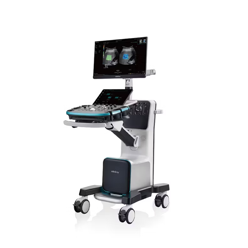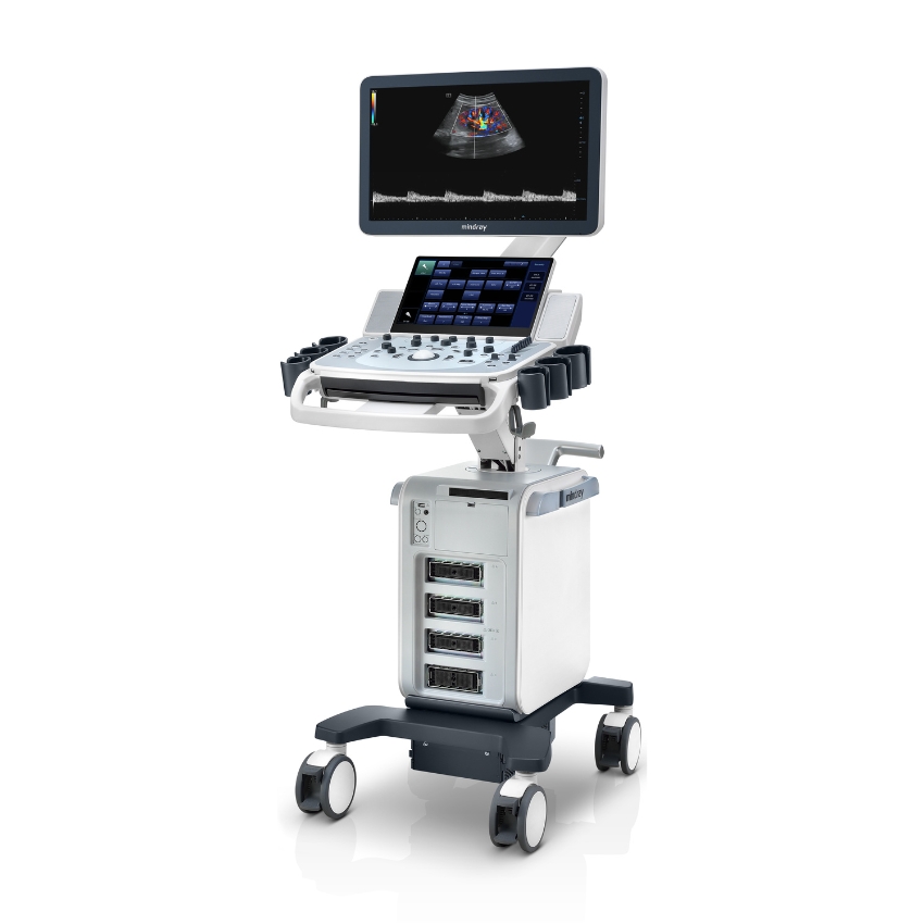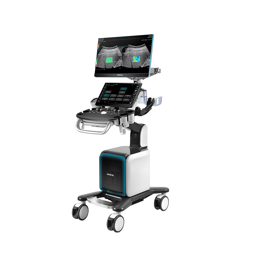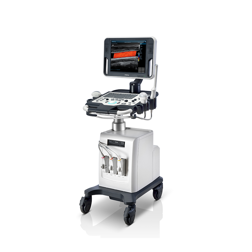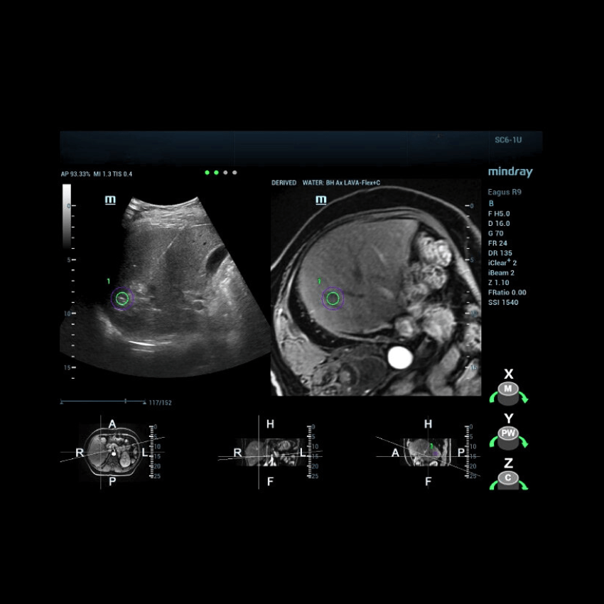
fusion imaging enables highly reliable lesion localization
can integrate CT, MRI, PET-CT, 3D ultrasound, and other imaging data with real-time 2D ultrasound
can also be used in ultrasound-guided interventional procedures
ideal when ultrasound alone cannot provide sufficiently accurate guidance for needle placement
features innovative respiratory compensation technology designed to reduce fusion distortion and inaccuracies caused by patient breathing
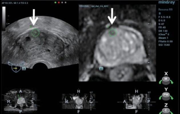
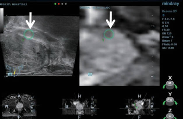
an important method for the diagnosis of prostate cancer (PCa)
can simulate ablation procedures, guide treatment, and provide three-dimensional information
Today, fusion biopsy is widely accepted in clinical ultrasound practice because it combines the advantages of CT/MRI imaging with real-time ultrasound, thereby improving the accuracy of both pathological diagnosis and ultrasound-guided interventions.
A commonly used analogy to explain the concept of fusion biopsy is a car GPS navigation system. Previously acquired CT/MRI data can be thought of as the map within the GPS system, while the real-time ultrasound image is comparable to what the human eye sees. By combining these two elements, the navigation system guides the “vehicle” — in this case, the ultrasound probe — to the desired location.
In general, every imaging modality has its own advantages and limitations.
Ultrasound is particularly valued for providing real-time imaging, avoiding ionizing radiation, and being relatively cost-effective compared with CT and MRI. However, because of its comparatively lower spatial resolution and the possibility of blind spots, certain lesions can sometimes be difficult to detect on ultrasound.
By contrast, the higher spatial resolution and wider field of view (FOV) provided by CT and MRI allow for more accurate lesion detection. However, CT and MRI examinations provide static images rather than real-time visualization, and CT additionally exposes the patient to ionizing radiation.
Fusion biopsy was therefore developed to combine the strengths of these imaging modalities while overcoming their individual limitations.
In addition to its diagnostic applications, fusion biopsy can also be used in ultrasound-guided interventional procedures.
A typical clinical scenario is ultrasound-guided radiofrequency ablation, particularly when ultrasound alone cannot provide sufficiently accurate guidance for needle placement. In such cases, by correlating the target lesion identified on MRI with the real-time ultrasound image, image fusion can accurately guide the needle tip to the lesion, thereby improving targeting precision.
Despite the clear advantages of fusion biopsy, unavoidable respiratory motion remains a major clinical challenge because it can reduce fusion accuracy. Clinical studies have shown that Mindray’s iFusion technology for fusion biopsy can correct approximately 80% of fusion errors caused by respiratory motion and significantly improve fusion accuracy throughout the respiratory cycle.
To address this clinical challenge, Mindray has developed an innovative and proprietary respiratory compensation technology supported by a highly sensitive magnetic motion sensor with millimeter-level precision. The technology is designed to minimize fusion distortion and inaccuracies caused by patient respiration.
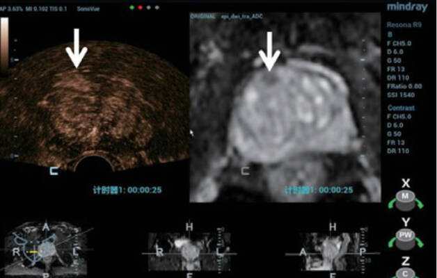
Compared with conventional fusion biopsy workflows, the new generation of iFusion with respiratory compensation requires only one additional step before the fusion navigation process. In addition to respiratory compensation, the widely recognized and highly valued Quad Mode provides simultaneous visualization of CT/MR image fusion with conventional tissue ultrasound and contrast-enhanced ultrasound. This makes it easier to visualize lesions across different imaging modalities. Such comprehensive, real-time multimodal imaging can provide greater diagnostic confidence.
In summary, combining the strengths of different imaging modalities offers significant benefits in everyday clinical practice, and Mindray continues to advance its healthcare solutions based on an in-depth understanding of key clinical needs.
Mindray’s innovative and patented iFusion technology delivers substantial clinical value by effectively reducing respiration-related artifacts. As a result, clinicians can approach the diagnosis of challenging lesions and interventional procedures with greater confidence, while benefiting from significantly improved fusion biopsy accuracy that takes image-guided procedures to a new level.
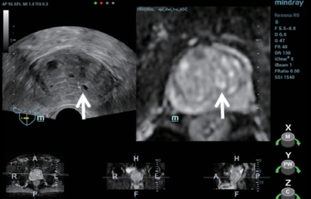
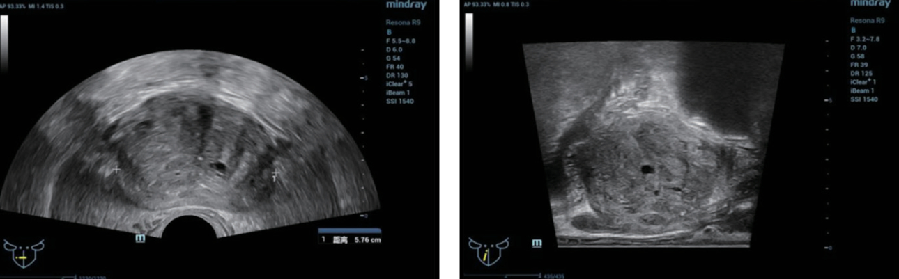
Figure 1 and 2. Conventional transrectal bi-planar ultrasound images of the prostate
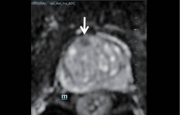
Figure 3. A low-signal lesion in the right anterior lobe of the prostate was most clearly visualized on the MRI ADC (apparent diffusion coefficient) sequence.
Figure 6. CEUS demonstrated early enhancement in the area corresponding to the lesion seen on MRI, compared with the contralateral peripheral tissue.
Figure 7. Visualization of the biopsy needle under iFusion guidance during sampling of the inferior portion of the lesion.
Send us an enquiry about the Mindray MR/CT/UZV iFusion and our team will get back to you with a quote, a demonstration date or an answer to your question.
Tell us how we can help. The selected offer is already attached to your enquiry.