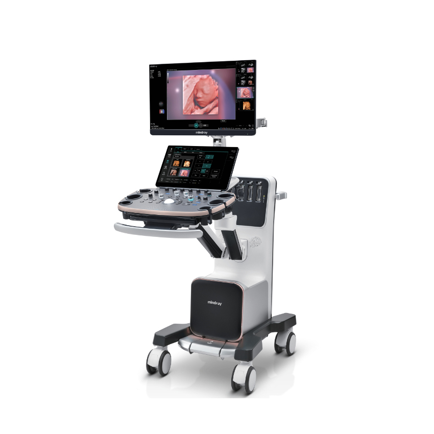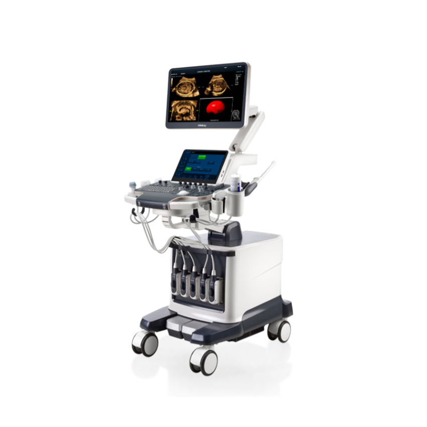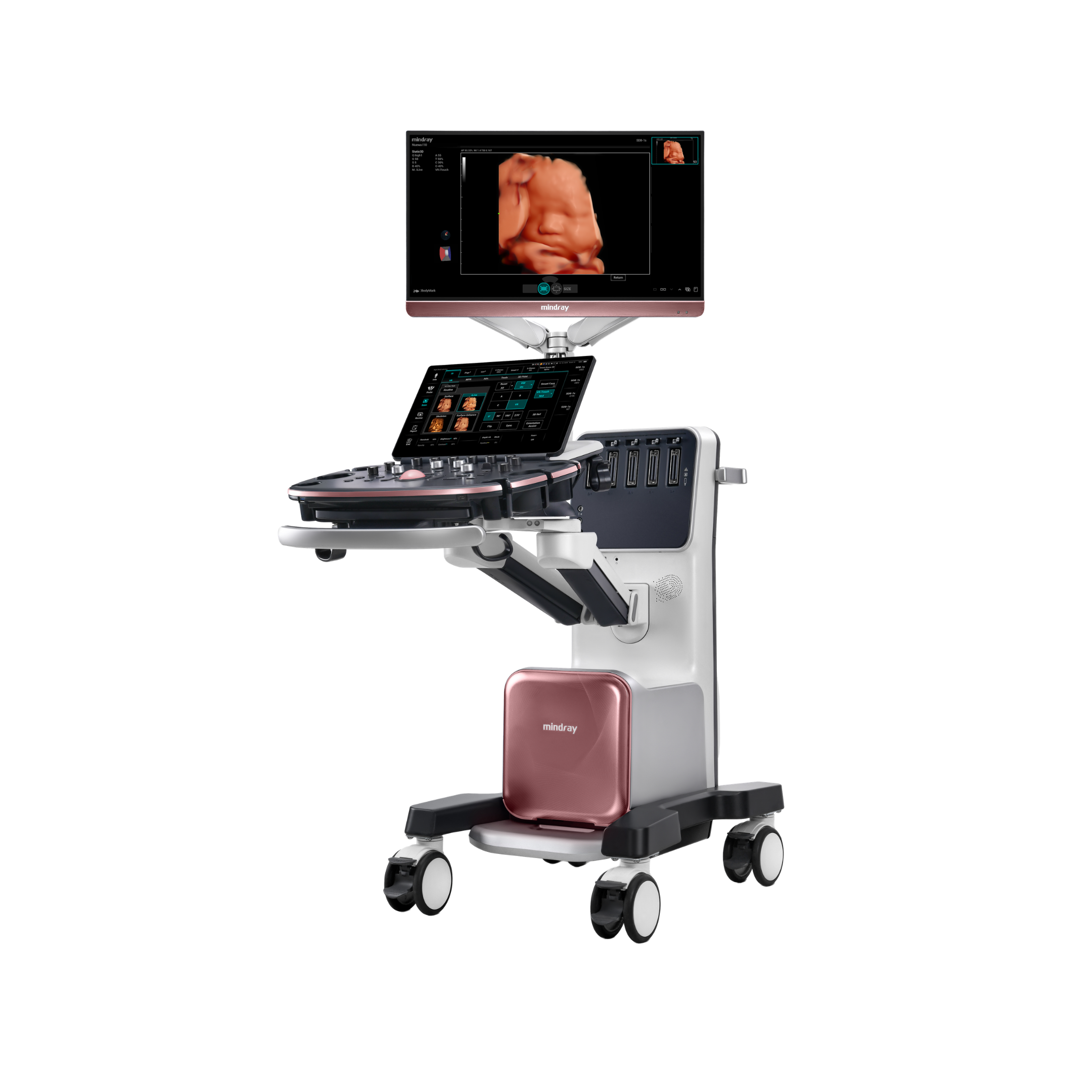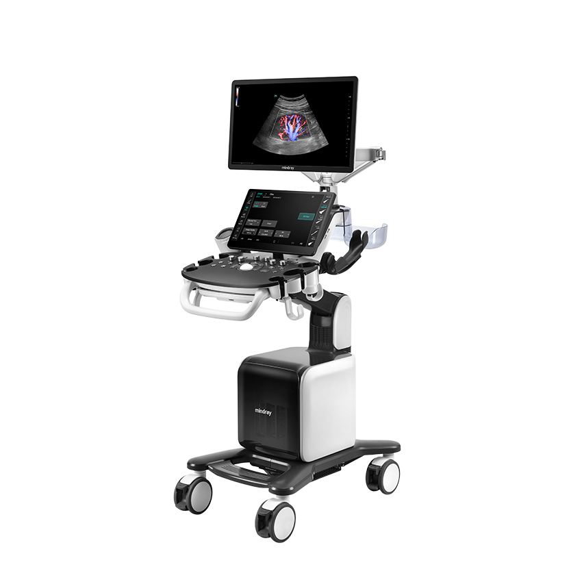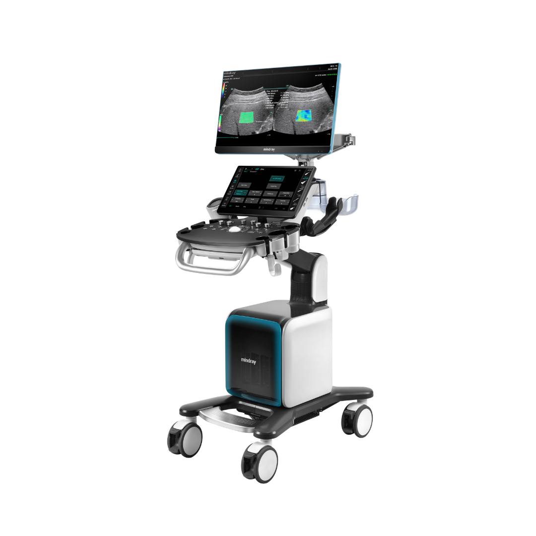
23.8” Full HD monitor and 15.6” touchscreen: the control panel is dust- and fluid-resistant
Applications: gynecology, obstetrics, fetal medicine, prenatal diagnosis, reproductive medicine and IVF, urogynecology
ZST+ technology: advanced ultrasound platform based on channel-data processing, designed to deliver high spatial and temporal resolution together with uniform tissue visualization
Advanced features: Smart Scene 3D, Smart Planes CNS, Smart Fetal HR, Smart Face, Smart OB, Smart FLC 2D/3D, iLive, Glazing Flow, HR Flow, as well as automated tools for fetal biometry, fetal central nervous system assessment, and reproductive medicine
Built-in battery: enables up to 90 minutes of scanning without connection to mains power

Mindray Nuewa N9 is an advanced ultrasound system designed primarily for gynecology, obstetrics, and reproductive medicine. It is based on the ZST+ platform, which, unlike conventional ultrasound signal processing, uses channel-data processing. This approach enables high-quality visualization of anatomical structures with good spatial and temporal resolution and uniform image quality.
The system combines advanced 2D, Doppler, and 3D/4D imaging with intelligent tools dedicated to women’s health. Smart Scene 3D combines volumetric imaging with automated recognition and analysis of anatomical structures, while Smart Planes CNS, Smart Face, and Smart OB simplify fetal anatomy assessment and biometric measurements. For reproductive medicine, Smart FLC 2D/3D provides automatic follicle detection, counting, and measurement. Realistic iLive volumetric rendering further improves visualization of fetal structures, while advanced Doppler technologies such as Glazing Flow and HR Flow provide more detailed visualization of vascularization and blood flow.
Mindray Nuewa N9 is an ultrasound system designed for routine gynecological and obstetric examinations, prenatal diagnosis, and monitoring of patients undergoing reproductive medicine procedures.
| Application areas |
Gynecology, obstetrics, fetal medicine, prenatal diagnosis, reproductive medicine and IVF, urogynecology |
|---|---|
| Imaging modes |
B-Mode, M-Mode, Color Doppler, Power Doppler, Pulsed Wave Doppler, CW Doppler, 3D imaging, real-time 4D imaging, volumetric imaging, and advanced Doppler imaging |
advanced platform that uses channel-data processing instead of conventional beamforming, enabling high spatial and temporal resolution together with uniform tissue visualization
monocrystalline transducer technology for improved penetration and more detailed imaging, particularly when examining deeper structures
transducer technology developed for increased imaging sensitivity and improved image quality
harmonic imaging for improved contrast and reduced artifacts
technology for reducing speckle noise and improving the clarity of anatomical structures
spatial compound imaging from multiple angles for improved contrast resolution and edge definition
high-resolution imaging for more detailed assessment of anatomical structures
advanced visualization of blood vessels and flow with a volumetric effect that facilitates assessment of vascular anatomy
sensitive Doppler imaging for visualization of small blood vessels and low-velocity blood flow
elastography technologies for assessment of tissue mechanical properties and stiffness
contrast-enhanced ultrasound imaging, depending on system configuration
real-time dynamic ultrasound image optimization
intelligent tools for automatic image optimization, measurements, and examination standardization
intelligent volumetric imaging developed to automate various steps of the obstetric examination
automatic recognition of standard fetal central nervous system planes and assistance with corresponding measurements
tool for fetal heart assessment and visualization
automatic one-click removal of structures obscuring the fetal face from the volumetric image
automated fetal biometry, enabling automatic measurement of commonly used obstetric parameters
automatic detection, counting, and measurement of follicles in 2D and 3D modes, intended for follicle assessment in reproductive medicine and IVF
realistic 3D/4D volumetric visualization of fetal structures
automatic recognition and display of standard anatomical planes
standardized examination protocol that guides the user through defined steps and helps reduce the possibility of omitting required views
built-in guide for performing examinations and learning ultrasound techniques
Send us an enquiry about the Mindray Nuewa N9 and our team will get back to you with a quote, a demonstration date or an answer to your question.
Probes selected for use with this ultrasound system.
Tell us how we can help. The selected offer is already attached to your enquiry.