NEW Portable ultrasound systems · mindray

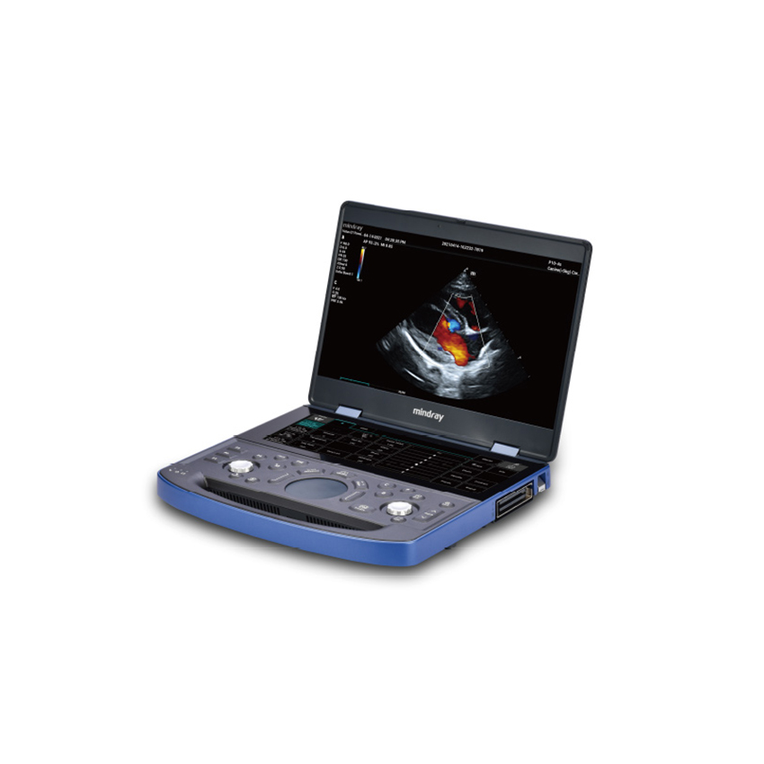
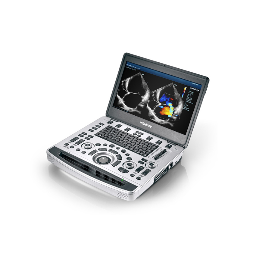
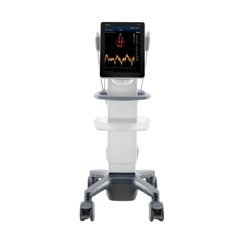
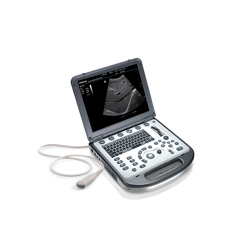
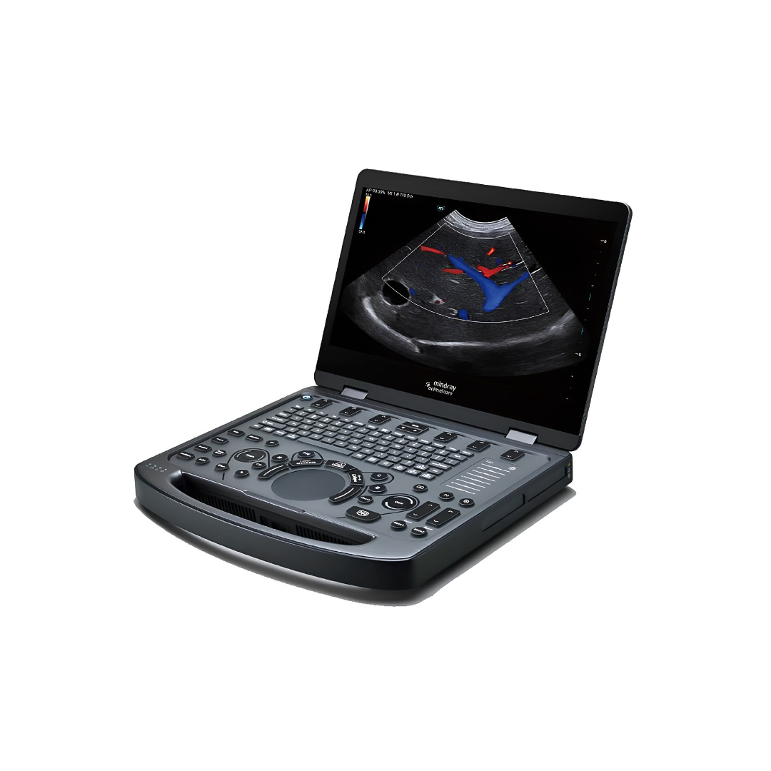
Mindray Vetus E3/E5

Portable Veterinary Ultrasound Systems for Everyday and Advanced Diagnostics

  • Applications: abdomen, reproduction, small parts, MSK, vascular examinations

  • 15.6” Full HD LCD display: resolution 1920 × 1080

  • Two probe ports

  • 1 TB SSD: large internal storage capacity

  • Weight: 3.5 kg, including battery

  • Li-ion battery: more than 1.5 hours of continuous scanning

  • Dimensions: 330 x 370 x 45 mm

  • Protected control panel: resistant to liquids, dust and animal hair; easy to clean and disinfect

Authorised distributor for Croatia · Installation, training and service included

Portable Ultrasound Systems Developed Specifically for Veterinary Practice

The Mindray Vetus E3 and Vetus E5 are compact color Doppler ultrasound systems designed for veterinary diagnostics. Their low weight, battery operation and compact dimensions make them suitable for use in veterinary clinics, hospitals and in the field, while the large 15.6-inch Full HD display provides excellent visualization of anatomical structures and blood flow. The built-in 1 TB SSD enables storage of a large number of examinations, while two probe ports allow quick switching between different clinical applications.

Both systems feature HR Flow, iClear, iTouch and Smart Track technologies to improve image quality and simplify everyday workflow. The Vetus E3 stands out with its broader cardiac package. It supports CW Doppler, Color M, anatomical M-mode, TDI and TDI QA for quantitative analysis of myocardial motion, including velocity, strain and strain rate. This makes it suitable for abdominal and general veterinary examinations, as well as more detailed echocardiographic diagnostics in dogs and cats.

The Vetus E5 focuses on advanced abdominal and soft-tissue diagnostics. Depending on the configuration, it supports UWN+ Contrast Imaging, Strain Elastography, Smart HRI, Smart Bladder, Smart 3D and iNeedle, enabling enhanced assessment of lesions, tissue elasticity, the liver and urinary bladder.

Technical specifications

Applications

Abdomen, cardiology, reproduction, small parts, vascular examinations and musculoskeletal examinations in dogs, cats, horses and other animal species

Image display modes

B-Mode, M-Mode, Color Doppler, Power Doppler, Pulsed Wave Doppler (PW), Continuous Wave Doppler (CW), Color M, duplex/triplex display, Tissue Harmonic Imaging (THI), Phase Shift Harmonic Imaging (PSH), Tissue Doppler Imaging (TDI), TDI QA, Free Xros M, Free Xros CM, iScape panoramic imaging and Smart 3D

Features

HR Flow

High-resolution visualization of microvascularization and complex blood-flow patterns; available in Color and Power Doppler modes

iClear

Intelligent image processing that reduces speckle noise, improves contrast and provides clearer tissue boundary definition

iTouch

Automatic optimization of ultrasound imaging parameters with a single touch

iBeam

Spatial compound imaging for improved image uniformity and visualization of anatomical structures

iZoom

Rapid enlargement of the ultrasound image and working area

iScape View

Panoramic imaging for visualization of an anatomical area larger than the probe's standard field of view

All features

Smart Track

Automatically optimizes the Color/Power Doppler region of interest as well as the position, angle and parameters of PW Doppler sampling

Smart Calc

Automatically recognizes the contour of a structure or lesion and performs the appropriate measurements and calculations

iWorks

Standardized, customizable examination protocols specifically designed for veterinary applications

iReport

Customizable veterinary reports with options for entering measurements and diagnostic descriptions, as well as saving and printing reports

iScanHelper

Built-in educational tool featuring reference ultrasound images, anatomical illustrations and probe-positioning guidance; on the E3, it includes more than 100 reference images and educational materials

1 TB SSD

Internal storage for examinations, images and videos

Wi-Fi and DICOM

Network connectivity, archiving and transfer of veterinary examinations

GalleryDrop

Easy transfer of images and videos to a mobile device using a QR code

Protected control surface

Enhanced resistance to liquids, dust and animal hair, allowing easier cleaning and disinfection

15.6" Full HD display

Resolution of 1920 x 1080, automatic brightness adjustment and opening angle of up to 180°

Compact design

Weight of 4 kg and housing dimensions of 330 x 370 x 45 mm

Battery operation

Replaceable rechargeable Li-ion battery providing 1.5 hours of continuous scanning

Key features

Mindray Vetus E3

TDI (Tissue Doppler Imaging)

Doppler analysis of myocardial motion for advanced cardiac examinations in dogs and cats

TDI QA

Quantitative analysis of myocardial motion velocity, strain and strain rate, with analysis of up to eight regions of interest

CW Doppler

Continuous-wave Doppler for measuring high flow velocities, particularly important in echocardiography

Free Xros M / Free Xros CM

Anatomical and curved anatomical M-mode for more flexible assessment of cardiac structures

Auto Calc

Automatic tracking of the Doppler spectrum and calculation of relevant parameters

Smart Track

Automatic tracking and optimization of Color, Power and PW Doppler imaging

Smart Calc

Automatic delineation and measurement of lesions and other structures

Key features

Mindray Vetus E5

Natural Touch / Strain Elastography

Assessment of the relative elasticity and stiffness of soft tissues as an additional aid in characterizing tissue changes

UWN+ Contrast Imaging

Highly sensitive contrast-enhanced ultrasound imaging for more detailed assessment of tissue vascularization and perfusion

Contrast Imaging QA

Analysis of time-intensity curves with the ability to work with multiple regions of interest

Smart HRI

Automatic identification of the liver and renal cortex and calculation of the hepatorenal index to support quantitative assessment of hepatic fatty infiltration

Smart Bladder

Automated assessment of the urinary bladder and bladder volume

iNeedle

Enhances needle visibility during biopsies, aspirations and other ultrasound-guided procedures

Smart 3D

Creation of a three-dimensional image based on manually acquired ultrasound data

Smart Calc

Automatic delineation of lesion boundaries and performance of multiple measurements

Request a quote or more information

Send us an enquiry about the Mindray Vetus E3/E5 and our team will get back to you with a quote, a demonstration date or an answer to your question.

Product Mindray Vetus E3/E5

We use your details only to answer this enquiry and never pass them on.

Related products

Scroll